Medical Image Segmentation
786 papers with code • 45 benchmarks • 44 datasets
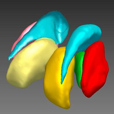
Medical Image Segmentation is a computer vision task that involves dividing an medical image into multiple segments, where each segment represents a different object or structure of interest in the image. The goal of medical image segmentation is to provide a precise and accurate representation of the objects of interest within the image, typically for the purpose of diagnosis, treatment planning, and quantitative analysis.
( Image credit: IVD-Net )

Benchmarks
These leaderboards are used to track progress in Medical Image Segmentation
| Trend | Dataset | Best Model | Paper | Code | Compare |
|---|---|---|---|---|---|

|
|||||

|
|||||

|
|||||

|
|||||

|
|||||

|
|||||

|
|||||

|
|||||

|
|||||

|
|||||

|
|||||

|
|||||

|
|||||

|
|||||

|
|||||

|
|||||

|
|||||

|
|||||

|
|||||

|
|||||

|
|||||

|
|||||

|
|||||

|
|||||

|
|||||

|
|||||

|
|||||

|
|||||

|
|||||

|
|||||

|
|||||

|
|||||

|
|||||

|
|||||

|
|||||

|
|||||

|
|||||

|
|||||

|
|||||

|
|||||

|
|||||

|
|||||

|
|||||

|
|||||

|
Libraries
Use these libraries to find Medical Image Segmentation models and implementationsDatasets
 DRIVE
DRIVE
 Kvasir-SEG
 GlaS
GlaS
 Kvasir
 Medical Segmentation Decathlon
 PROMISE12
 AMOS
AMOS
 CHASE_DB1
 CVC-ClinicDB
 ACDC
ACDC
Subtasks
-
 Lesion Segmentation
-
 Brain Tumor Segmentation
-
 Cell Segmentation
-
Skin Lesion Segmentation
-
Skin Lesion Segmentation
-
 Brain Segmentation
-
Semi-supervised Medical Image Segmentation
-
 Retinal Vessel Segmentation
-
MRI segmentation
-
Cardiac Segmentation
-
 3D Medical Imaging Segmentation
-
Liver Segmentation
-
Volumetric Medical Image Segmentation
-
Brain Image Segmentation
-
 Pancreas Segmentation
-
Iris Segmentation
-
Video Polyp Segmentation
-
Lung Nodule Segmentation
-
Nuclear Segmentation
-
COVID-19 Image Segmentation
-
Skin Cancer Segmentation
-
Electron Microscopy Image Segmentation
-
Ischemic Stroke Lesion Segmentation
-
Brain Lesion Segmentation From Mri
-
Placenta Segmentation
-
Infant Brain Mri Segmentation
-
Automatic Liver And Tumor Segmentation
-
Acute Stroke Lesion Segmentation
-
Cerebrovascular Network Segmentation
-
Automated Pancreas Segmentation
-
Semantic Segmentation Of Orthoimagery
-
Pulmorary Vessel Segmentation
-
Brain Ventricle Localization And Segmentation In 3D Ultrasound Images
Most implemented papers
UNet++: Redesigning Skip Connections to Exploit Multiscale Features in Image Segmentation
The state-of-the-art models for medical image segmentation are variants of U-Net and fully convolutional networks (FCN).
UNETR: Transformers for 3D Medical Image Segmentation
Project-MONAI/research-contributions
•
 •
•
Inspired by the recent success of transformers for Natural Language Processing (NLP) in long-range sequence learning, we reformulate the task of volumetric (3D) medical image segmentation as a sequence-to-sequence prediction problem.
nnU-Net: Self-adapting Framework for U-Net-Based Medical Image Segmentation
MIC-DKFZ/nnunet
•
•
The U-Net was presented in 2015.
Uncertainty-aware Self-ensembling Model for Semi-supervised 3D Left Atrium Segmentation
yulequan/UA-MT
•
•
We design a novel uncertainty-aware scheme to enable the student model to gradually learn from the meaningful and reliable targets by exploiting the uncertainty information.
Automatic Brain Tumor Segmentation using Cascaded Anisotropic Convolutional Neural Networks
charan223/Brain-Tumor-Segmentation-using-Topological-Loss
•
 •
•
A cascade of fully convolutional neural networks is proposed to segment multi-modal Magnetic Resonance (MR) images with brain tumor into background and three hierarchical regions: whole tumor, tumor core and enhancing tumor core.
A Novel Focal Tversky loss function with improved Attention U-Net for lesion segmentation
nabsabraham/focal-tversky-unet
•
•
We propose a generalized focal loss function based on the Tversky index to address the issue of data imbalance in medical image segmentation.
MultiResUNet : Rethinking the U-Net Architecture for Multimodal Biomedical Image Segmentation
nibtehaz/MultiResUNet
•
•
ScienceDirect 2019
We have compared our proposed architecture MultiResUNet with the classical U-Net on a vast repertoire of multimodal medical images.
ResUNet++: An Advanced Architecture for Medical Image Segmentation
Accurate computer-aided polyp detection and segmentation during colonoscopy examinations can help endoscopists resect abnormal tissue and thereby decrease chances of polyps growing into cancer.
UNet 3+: A Full-Scale Connected UNet for Medical Image Segmentation
UNet, which is one of deep learning networks with an encoder-decoder architecture, is widely used in medical image segmentation.
Boundary loss for highly unbalanced segmentation
We propose a boundary loss, which takes the form of a distance metric on the space of contours, not regions.
